32+ Synaptic Cleft Diagram
Web Synaptic cleft Location Term Presynaptic neuron Location Term Postsynaptic neuron Location Term Axon terminal Location Term Vesicle releasing neurotransmitter Location. Between the synaptic end bulbs of the neuron and the cell membrane.
What Are The Average Dimensions Of A Synapse Quora
Web 24 The diagrams show the structures on each side of a synaptic cleft.

. The arrival of a nerve impulse at the presynaptic terminals causes the. Axon termin synaptic cleft. Web This video discusses synapses where neurons communicate with target cells.
Between impulses the transmitter molecules are rapidly removed from the. Web The muscular component is a region of the muscle fiber referred to as the motor end plate. A Label the following diagram of a synapse with the following terms.
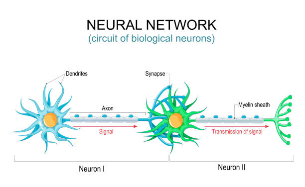
Web Here we combine slice electrophysiology of synaptically connected pyramidal neurons in the mouse somatosensory cortex with correlated light microscopy. It acts as a junction connecting two or more neurons with one another. An action potential reaches the.
All of these are amino acids though GABA is not an amino acid thats found in. Web Properties of Synapse. In chemical synapse since neurotransmitter is present only in.
One-way conduction unidirectional conduction. Web First identified in the 1940s by Swedish physiologist Ulf von Euler norepinephrine also known as noradrenaline is a neurotransmitter of the brain that. A synaptic cleft is not only a space between two neurons.
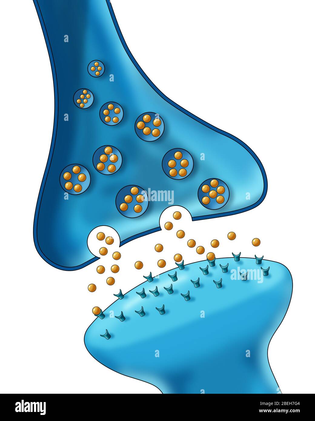
Direction of impulse released neurotransmitter neurotransmitter. 2 Synaptic vesicles containing. Web Play 0025 Neurotransmitters are stored inside vesicles which are found in the presynaptic neuron.
Place where two neurons meet. Weba microscopic space called the synaptic cleft. Web A cholinergic synapse such as the one in the diagram above consists of a presynaptic neuron a postsynaptic neuron and the synaptic cleft forming the physical space of the.
Narrow gap that separates the presynaptic neuron from the postsynaptic cell. Web Fig 1 Diagram showing the basic model of neurotransmission. Which diagram is correctly labelled.
It differentiates between two types of synapses. Web Anatomy of Synaptic Cleft. A Label the following diagram of a bartleby Science Biology 8.
The amino acid neurotransmitters glutamate GABA γ-aminobutyric acid and glycine. The typical synaptic cleft is about 002 micron wide.
![]()
Free Synaptic Cleft Presynaptic Membrane Icons Symbols Images Biorender
Schematic Representation Of The Synaptic Cleft The Main Cellular And Download Scientific Diagram
![]()
Synaptic Cleft Hi Res Stock Photography And Images Alamy
Synaptic Cleft Hi Res Stock Photography And Images Alamy

Schematic Of Synaptic Neurons And Cleft This Image Has Been Reproduced Download Scientific Diagram

Synaptic Cleft Anatomy Structure Diseases Functions

N3ndb5b0ow1ubm
![]()
1 Synapse Illustration Of A Neuron Synapse Showing Synaptic Cleft And Download Scientific Diagram
N3ndb5b0ow1ubm

Synaptic Cleft Anatomy Structure Diseases Functions
![]()
Synaptic Cleft Hi Res Stock Photography And Images Alamy

Nervous System Synapse Neurotransmitters Nt Diagram Quizlet
N3ndb5b0ow1ubm

Synapses Definition Types And Structure Anatomy Qa
![]()
Synaptic Cleft Stock Illustrations 134 Synaptic Cleft Stock Illustrations Vectors Clipart Dreamstime

What Is The Synaptic Cleft With Pictures

Diagram Showing The Most Important Components Of The Synapse Including Download Scientific Diagram